










For over twelve years, he had lived with chest pain on exertion, which he attributed to anxiety and gastric reflux . Then, a few weeks ago, he arrived at the emergency room of Carbonia hospital for acute coronary syndrome: severe chest pain, changes in his electrocardiogram, and the fear of a heart attack.
Behind those symptoms, however, lay a rare and complex pathology: a giant coronary fistula , a congenital anomaly that creates an abnormal communication between the coronary arteries and the pulmonary artery, altering the normal blood flow to the heart and causing myocardial ischemia.
The patient, a 67-year-old woman, was immediately transferred to Brotzu Hospital in Cagliari, where the Pediatric Cardiology and Congenital Heart Disease team, led by Dr. Angelica Rossi, completed diagnostic tests, ruling out the presence of any other congenital heart malformations.
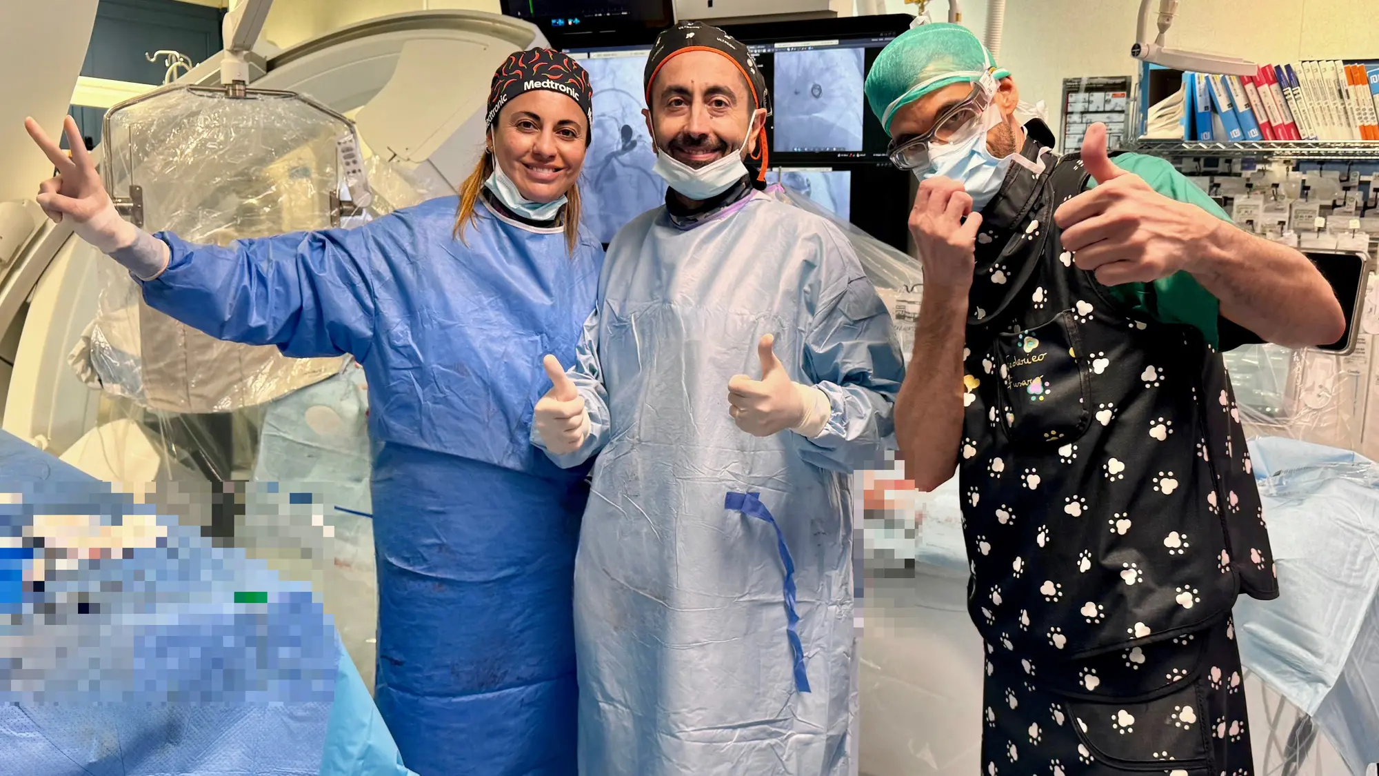
On May 7, 2026, the woman underwent a highly complex, minimally invasive procedure , performed without opening the chest and without general anesthesia. This delicate and highly specialized procedure was completed thanks to the combined efforts of various professionals, integrating cardiology, hemodynamics, and neuroradiology expertise.
The surgery was performed by doctors Angelica Rossi , director of the Congenital Interventional Cardiology unit, Federico Fusaro , director of Interventional and Vascular Neuroradiology, and Alberto Boi of Interventional and Haemodynamic Cardiology.
"To close the fistula," Brotzu reports, "an innovative method was used: twelve electrolyte-releasing microcoils were inserted into the aneurysm sac and released one at a time through a controlled detachment system using low-voltage current. This technology was used for the first time in this type of cardiac surgery . The coils, folding inside the fistula, interrupted the abnormal blood flow, causing thrombosis and allowing blood to return to the heart's proper flow, rather than leaking into the pulmonary artery."